EXo-i
세포 배양액과 혈액으로부터신속하게 엑소좀을 분리 할 수 있습니다.
Exo-i로 수용성 단백질과 지질 단백질을효과적으로 제거하여 고순도의 엑소좀을 획득하세요.
칼럼크로마토그래피 방법을 이용하여, 엑소좀에 다른 화학적, 물리적 변형 없이 입자 크기에 따른 경로차이를 이용하여
고순도의 엑소좀을 획득할 수 있어 후속 연구에 활용이 용이합니다.
세포 배양액과 혈액으로부터신속하게 엑소좀을 분리 할 수 있습니다.
Exo-i로 수용성 단백질과 지질 단백질을효과적으로
제거하여 고순도의 엑소좀을 획득하세요.
칼럼크로마토그래피 방법을 이용하여,
엑소좀에 다른 화학적, 물리적 변형 없이 입자 크기에 따른
경로차이를 이용하여 고순도의 엑소좀을 획득할 수 있어
후속 연구에 활용이 용이합니다.
Why EXo-i
-
- 안전한 분리
- 엑소좀 손상없이 분리
-
- 빠른 분리
- 15분 이내의 엑소좀 분리
-
- 고순도 분리
- 효과적인 수용성, 지질 단백질 제거
기술원리
다중층 크기배제 크로마토그래피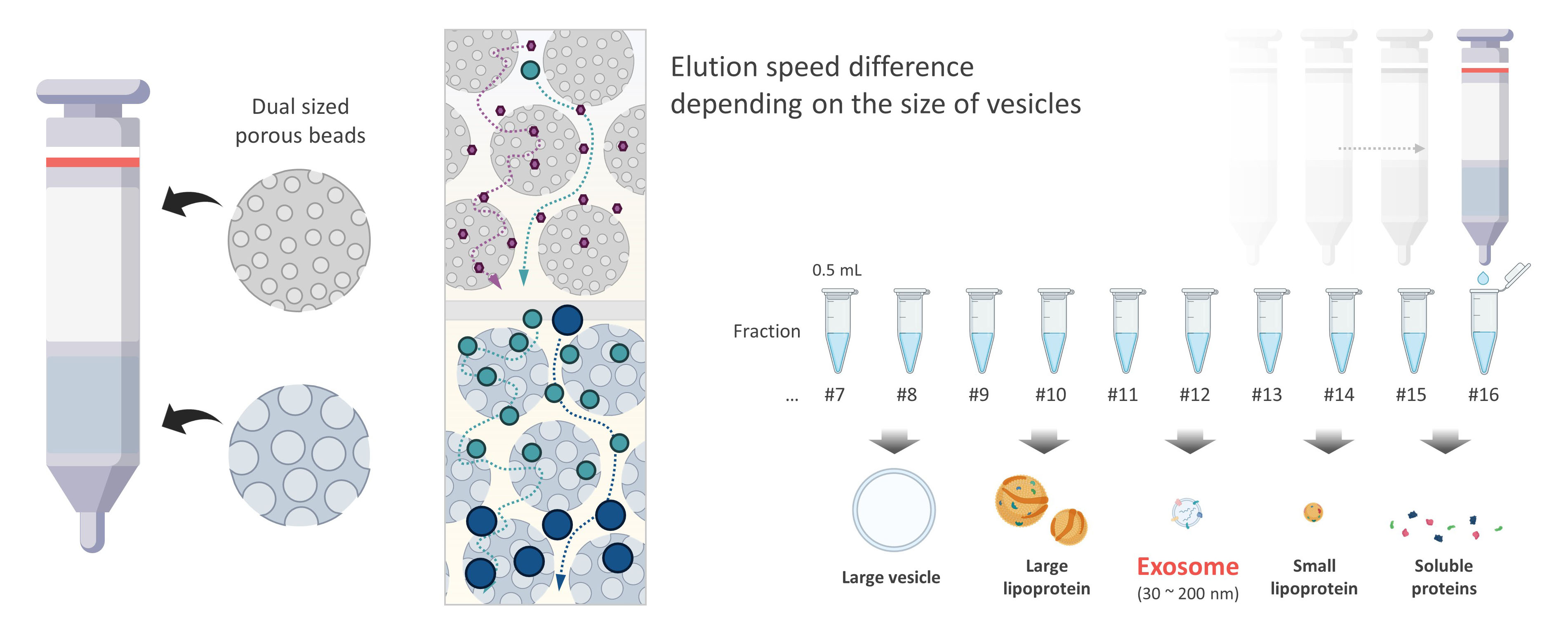
Exo-i는 칼럼 내부에 채워진 두개층의 다공성 비드를 통해 엑소좀을 분리할 수 있습니다.
각각의 비드는 서로 다른 공극을 가지고 있으며 복합적인 크기 배제를 통해 엑소좀 분획을 획득하는 것을 넘어
혈장 분석에서 간섭을 일으키는 지질단백질 및 수용성 단백질을 효과적으로 제거할 수 있습니다.
(관련 논문: Scientific reports 11.1 (2021): 1-9.)
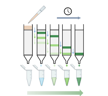
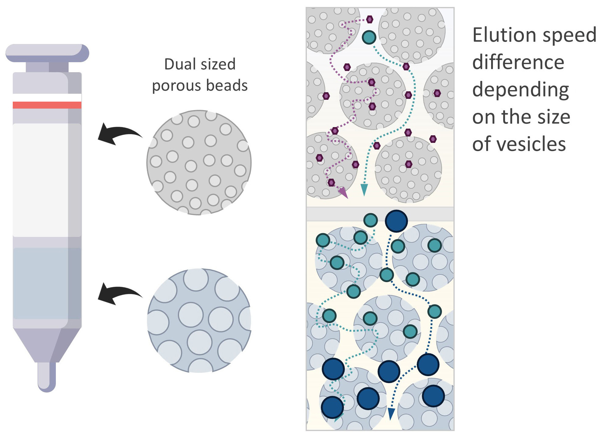
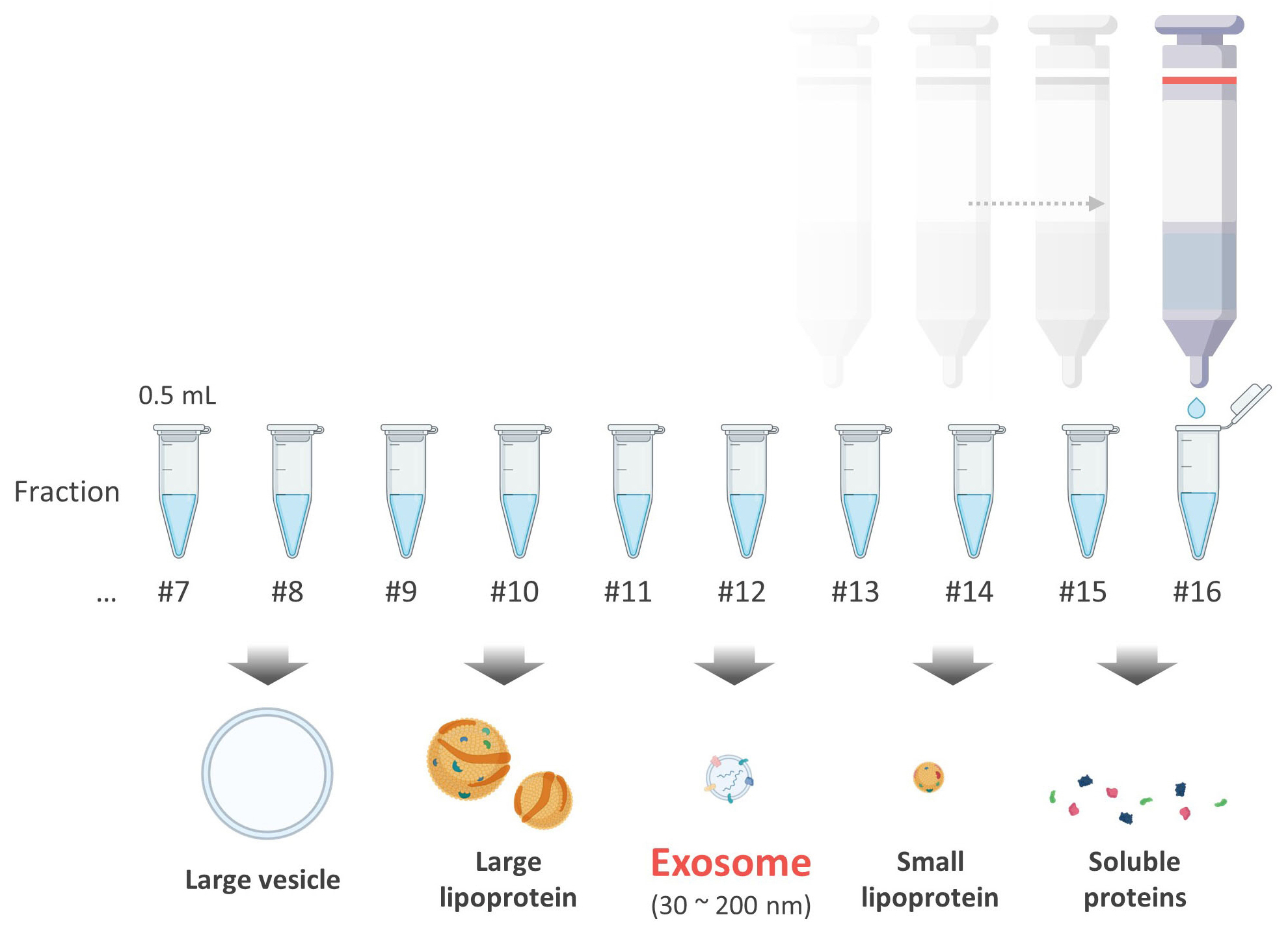
- Exo-i를 이용하는 방법은
매우 단순합니다. -
- 칼럼에 PBS를 흘려주고 분리를 준비합니다.
- 생체샘플(세포배양액, 혈장, 혈청 등) 500 ㎕를 로딩합니다
- 물방울이 떨어지지 않으면 PBS를 추가합니다.
- 분획을 500 ㎕ 씩 포집합니다.
성능결과
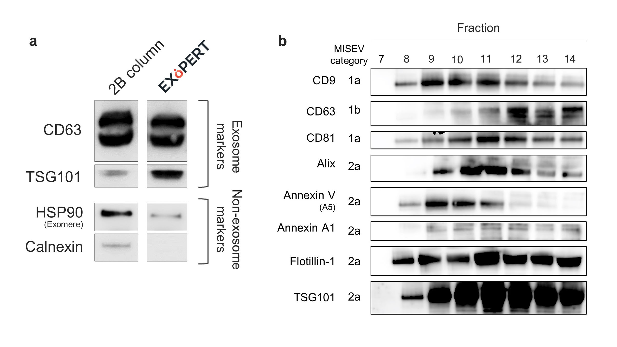
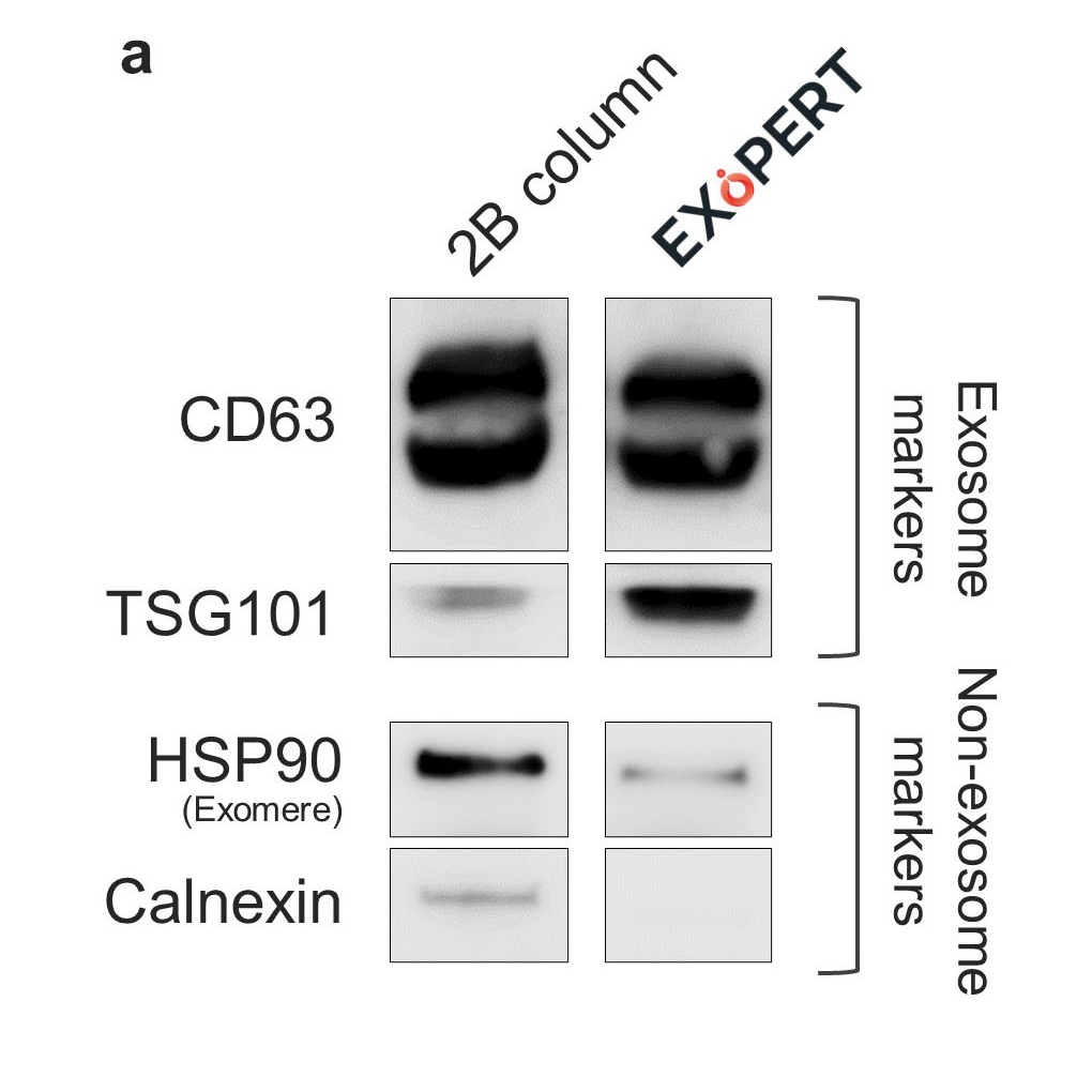
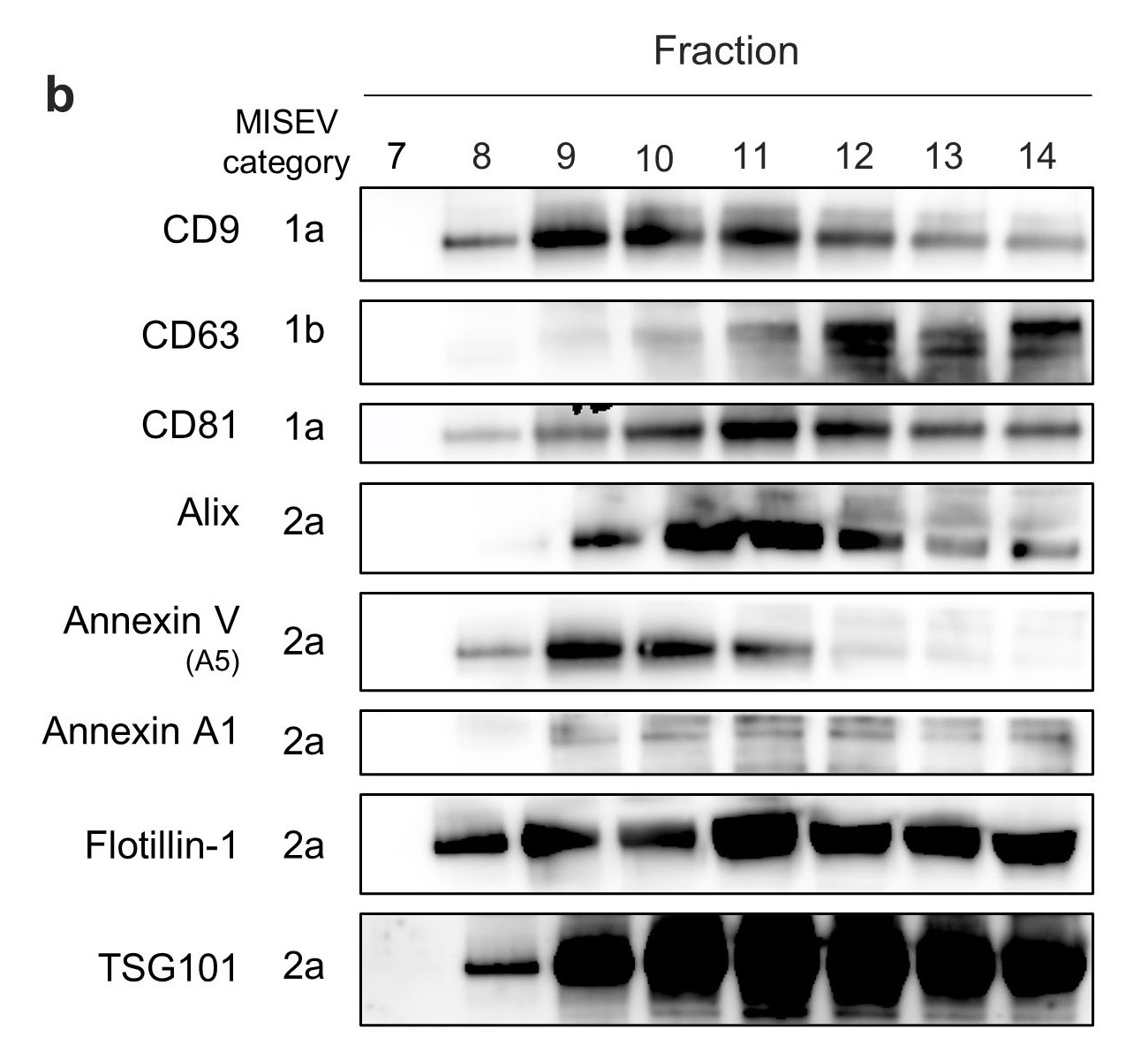
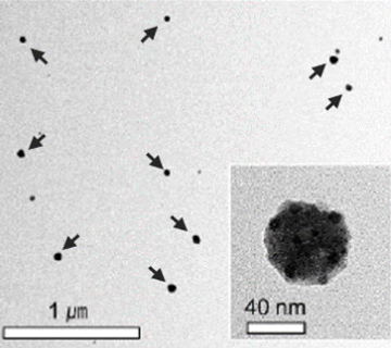
- Exosomal protein expression
- Western blotting (WB) results to show EV markers from a) lung cancer cell media and b) human plasma. a) The result shows more effective removal of non-exosome markers and preservation of exosome markers compared to a conventional 2B column. b) The result indicates protein expression of exosomal markers (CD9, CD63, CD81, Alix, Annexin V, Annexin A1, Flotillin-1, and TSG101 by fractions. The decision on the type of protein markers followed the MISEV2018 guidelines.
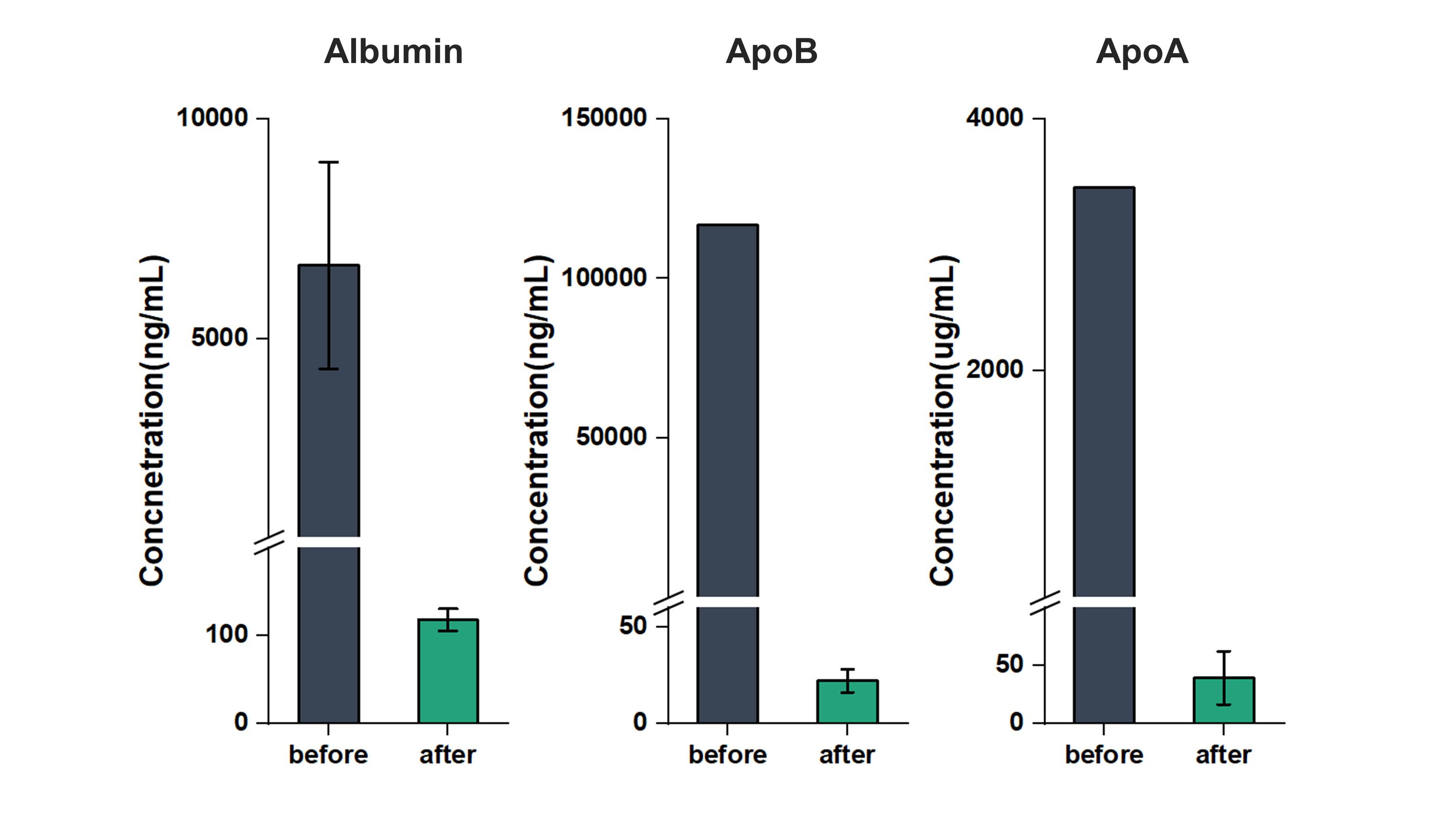
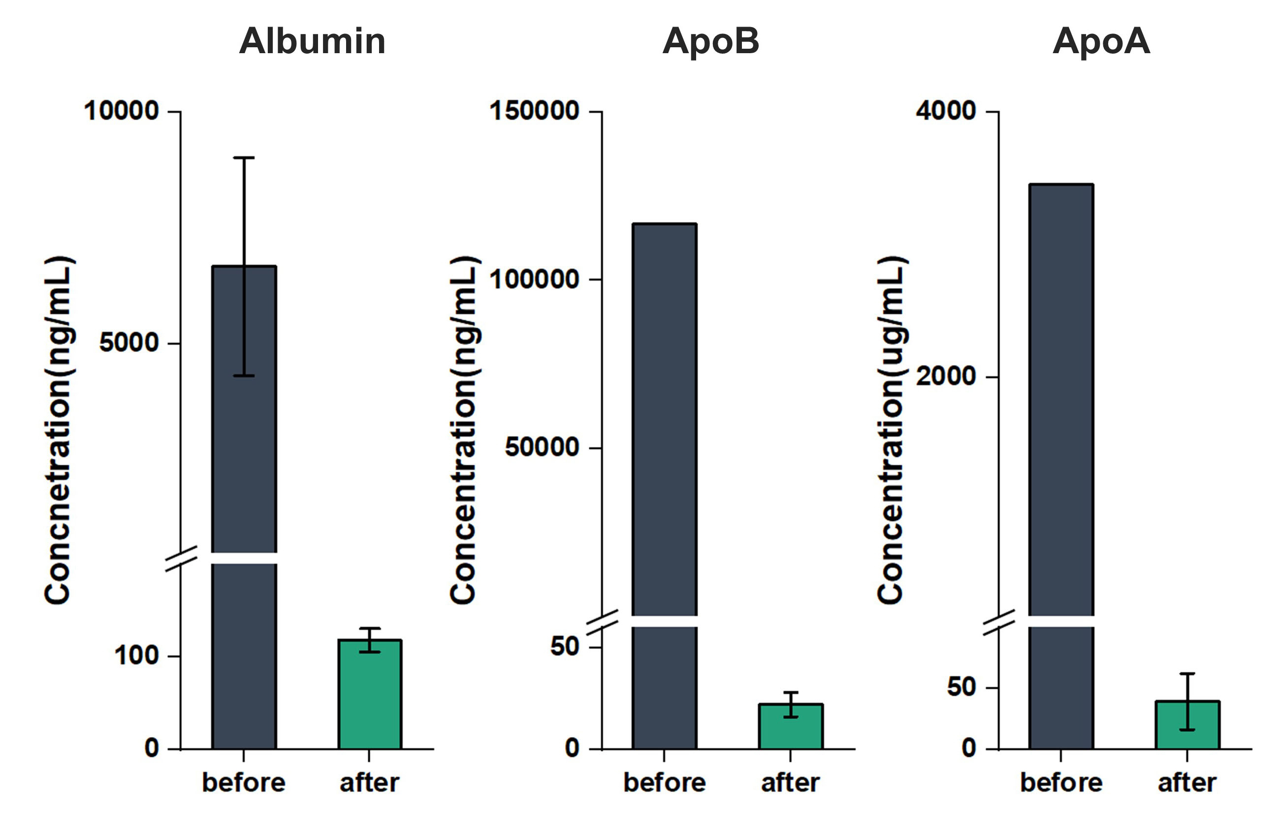
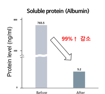
- Elimination of lipoprotein and
soluble proteins from plasma - Removal proportion of lipoproteins and soluble proteins from plasma. The concentration was measured using Enzyme-Linked Immunosorbent Assay (ELISA). The analysis shows that albumin decreased by 91.1%, ApoB by 92.6%, and ApoA by 92%.
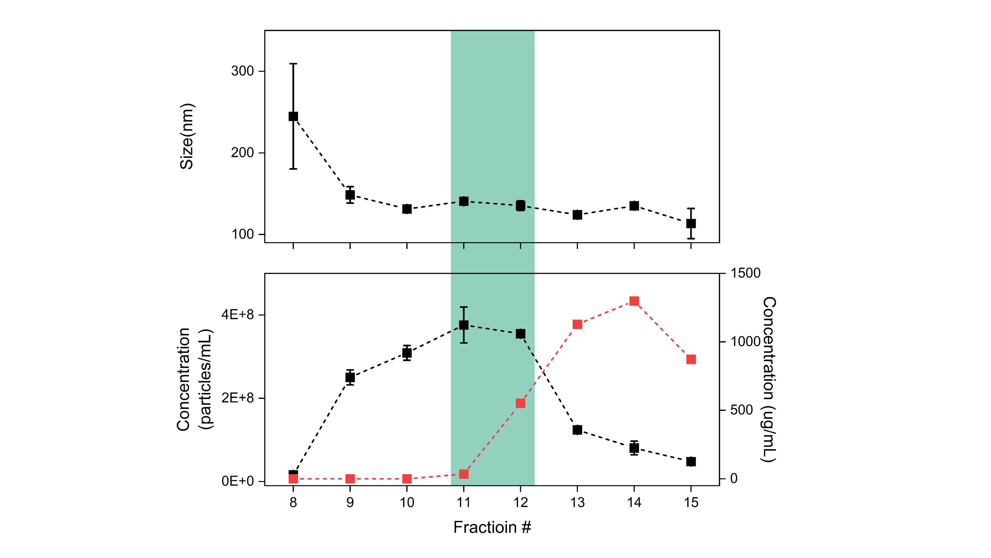
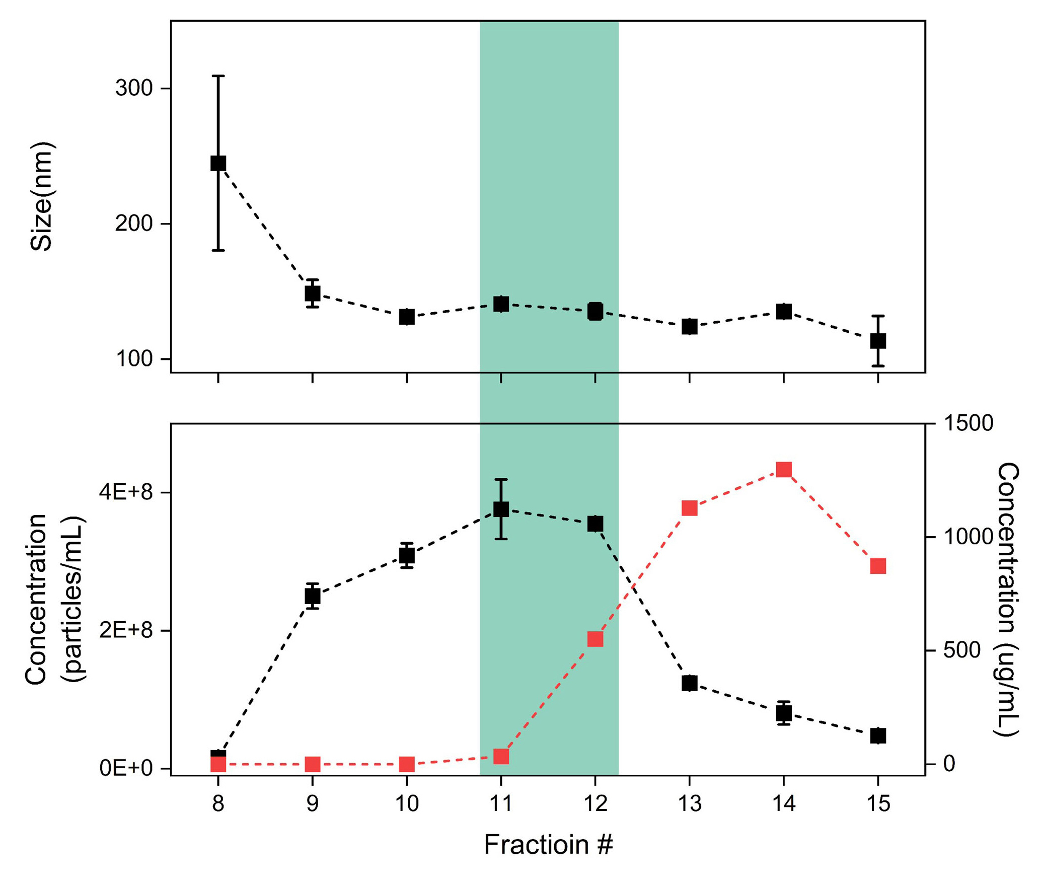
- Size, yield, and purity
- Exosome size distribution and yields from plasma by nanoparticle tracking analysis (NanoSight) and small protein distribution. The size distribution shows a gradual decrease in vesicle size according to fractionation. The mode sizes at fractions 11 and 12 are in the range of 100 ~ 150 nm. The bottom graphs show the concentration distribution of vesicle-like particles (black) and small proteins (red). This result suggests that our method can avoid interference by free protein as much as possible. The particle concentrations are about 5.0 × 10⁸ particles/mL at fractions 11 and 12.
논문 및 관련 문서
Many papers have been published using our technology.
- "Early-stage lung cancer diagnosis by deep learning-based spectroscopic analysis of circulating exosomes."
ACS nano 14.5 (2020): 5435-5444.
- "A Proteomic Approach to Understand the Clinical Significance of Acute Myeloid Leukemia–Derived Extracellular Vesicles Reflecting Essential Characteristics of Leukemia."
Molecular & Cellular Proteomics 20 (2021).
- "Dual size-exclusion chromatography for efficient isolation of extracellular vesicles from bone marrow derived human plasma."
Scientific reports 11.1 (2021): 1-9.
- "GCC2 as a New Early Diagnostic Biomarker for Non-Small Cell Lung Cancer."
Cancers 13.21 (2021): 5482.